Pigmented Oral Lesions in Thailand
The most prevalent pigmented oral lesion in Thailand is nevus, suggests a recent study published in the European Journal of Dentistry. The aim of this study was to determine the prevalence and clinical features of pigmented oral lesions in Thailand.
Methodology
Biopsy records of the following departments were reviewed for oral pigmented lesions diagnosed during 1999 to 2019:
- Department of Oral Pathology, Chulalongkorn University
- Department of Oral Diagnosis, KhonKaen University
- Department of Oral Biology and Oral Diagnostic Sciences, Chiangmai University
- Department of Stomatology, Prince of Songkla University
- Rangsit University
Demographic data were culled from the biopsy records. The ages of the patients were subdivided into 10-year intervals. Locations of the lesions were classified as gingiva, labial/buccal mucosa, palate, the floor of the mouth, tongue, as well as a combination of sites. Data were analyzed by descriptive statistics using SPSS version 20.0.
Results
- Of the 47,175 accessioned cases, 241 cases (0.51%) were diagnosed in the category of pigmented oral lesions.
- The age of the patients ranged from 1 month to 88 years with the mean ± standard deviation = 38.74 ± 20.96 years.
- Regarding gender, 172 patients (71.37%) with pigmented lesions were females, while 69 patients (28.63%) were males.
- The female-to-male ratio was 2.49:1.
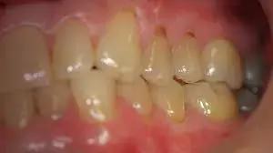
- The majority of the pigmented lesions were encountered at the gingiva (29.88%) followed by labial/buccal mucosa (26.97%), palate (14.94%), lip (10.79%), alveolar mucosa (9.54%), and others (7.88%), respectively.
- The three most common pigmented oral lesions in the present study were nevus (39.83%), followed by melanotic macule (28.63%) and amalgam tattoo (17.43%), respectively.
Thus, the most common pigmented oral lesion in the present study is nevus. Demographic data of the patients in the present study are in accordance with previous studies with minor differences. Even though pigmented lesions of the oral cavity constitute a small portion of the oral pathology biopsies, accurate diagnosis is important since there is an overlap in the clinical appearance of benign pigmented lesions and melanoma.
Reference
Kittipong Dhanuthai et al. Pigmented Oral Lesions: A Multicenter Study. CC BY 4.0 · Eur J Dent 2022; 16(02): 315-319. DOI: 10.1055/s-0041-1735790
0 Comments
Post a comment
No comments yet. Be the first to comment!