Ocular Imaging May Provide Early Information On Microvascular Damage In PAD Patients
Peripheral artery disease (PAD) is a prevalent vascular disorder associated with atherosclerosis and reduced blood flow to the extremities. Affecting 8-10 million Americans and over 200 million people globally, it is a complex condition influenced by various risk factors like diabetes, smoking, hypertension, high cholesterol, and coronary artery disease. PAD leads to structural and functional changes in the limbs, resulting in disability, decreased quality of life, increased mortality, etc.
A recent study published in Vascular Medicine concluded that a more specific biomarker for PAD is necessary due to the poor predictive value of clinical assessment. A noninvasive ophthalmic imaging technique can significantly improve the diagnosis rate without putting the patient at risk for complications. Early detection and treatment of PAD can enhance quality of life and improve clinical outcomes.
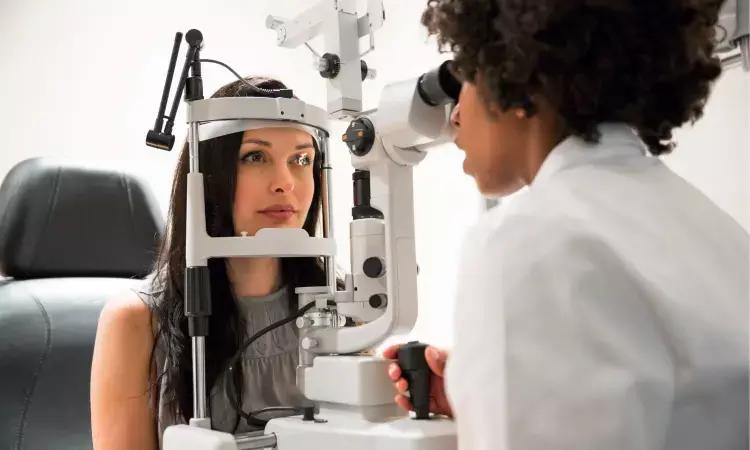
This study reviewed the current literature on noninvasive ocular imaging for the PAD diagnosis. The search included Medline, Scopus, Embase, Cochrane, and other databases, with five articles selected. Two studies used retinal colour fundus photography, one used optical coherence tomography (OCT), and two used optical coherence tomography angiography (OCTA) to assess ocular changes in PAD.
Key findings from the study are:
· PAD patients showed both structural and functional changes in the retina.
· Structural alterations were found around PAD patients' optic disc and temporal retinal vascular arcades.
· Retinal hemorrhages, exudates, and microaneurysms, detected through color fundus photography, were associated with an increased future risk of PAD.
· The RNFL (retinal nerve fibre layer) was thinner in the nasal quadrant of PAD patients compared to age-matched healthy individuals in OCT.
· choroidal thickness in the subfoveal region was thinner in PAD patients controls.
· OCTA revealed a significant reduction in the retinal and choroidal circulation in PAD patients compared to healthy controls.
As PAD causes thinning and ischemic changes in retinal vessels, retinal imaging techniques can provide valuable information about early microvascular damage in PAD. Ocular imaging may serve as a potential biomarker for PAD.
Study limitations include a smaller number of studies, bias, only two studies using OCTA, and none of the studies investigating the association between retinal changes and disease severity.
Reference:
Prem Senthil et al. Role of noninvasive ocular imaging as a biomarker in peripheral artery disease: A systematic review. Vascular Medicine. 2023;0(0).
Disclaimer: This website is designed for healthcare professionals and serves solely for informational purposes.
The content provided should not be interpreted as medical advice, diagnosis, treatment recommendations, prescriptions, or endorsements of specific medical practices. It is not a replacement for professional medical consultation or the expertise of a licensed healthcare provider.
Given the ever-evolving nature of medical science, we strive to keep our information accurate and up to date. However, we do not guarantee the completeness or accuracy of the content.
If you come across any inconsistencies, please reach out to us at
admin@doctornewsdaily.com.
We do not support or endorse medical opinions, treatments, or recommendations that contradict the advice of qualified healthcare professionals.
By using this website, you agree to our
Terms of Use,
Privacy Policy, and
Advertisement Policy.
For further details, please review our
Full Disclaimer.
0 Comments
Post a comment
No comments yet. Be the first to comment!